
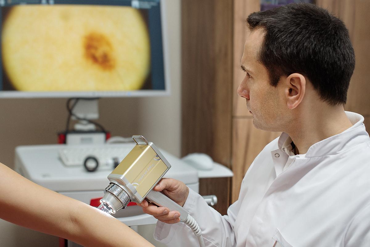
Regular videodermatoscopy examinations are the most effective method for the early detection of melanoma and other skin cancers.
Why is it worth it?
Digital monitoring of changes over time
- Image archiving: The device stores images in the patient’s database.
- Comparison: At each follow-up visit, the doctor compares previous images with current ones.
- Detecting evolution: The system immediately highlights even minimal changes in a mole’s structure, size, or colour.
Precision and advanced technology
- High magnification: The digital camera magnifies the image 15 to as much as 70–100 times, while a classic dermatoscope magnifies only 10 times.
- Body mapping: Advanced systems create a photographic map of the whole skin, making new moles easier to locate.
- AI support: Modern software uses artificial intelligence for preliminary risk analysis.
Safety and avoiding unnecessary procedures
- Fewer excisions: A thorough assessment helps avoid the preventive removal of benign lesions.
- Painless: The examination is 100% non-invasive and comfortable for the patient.
- No restrictions: It can be safely repeated in children and pregnant women.
Who should be screened most often?
A preventive examination should be performed at least once a year. Based on the examination, your doctor will advise how often you should return for follow-up.
If a lesion has grown or changed its appearance, it is best to consult a doctor.